
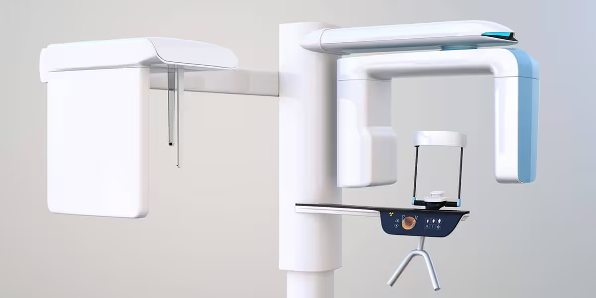
3D Cone Beam Imaging: A Clearer, Safer View of Your Teeth and Jaw
Cone Beam technology provides a digital tomographic 3D view of the patient’s area of interest. A traditional x-ray is only two-dimensional. With Cone Beam systems the doctor is able to get a full 360-degree view of the tooth and all surrounding areas. The 3D Cone Beam scanner provides nearly limitless views of the teeth while using less radiation than traditional medical CT technology. This new technology is fast, simple, and painless, providing many wonderful benefits that were unavailable only a few years ago.
Click here to get a list of phone numbers and schedule an appointment in one of our readily available offices today!
How CBCT Compares on Radiation Exposure
Radiation safety is a reasonable question to ask about any imaging procedure, and the peer-reviewed literature offers a fairly clear answer for CBCT. Dental CBCT uses a pulsed, narrowly focused beam and a single rotation around the patient, which produces a dramatically smaller dose than a conventional medical CT of the maxillofacial area. One study published in the Journal of Pharmacy and Bioallied Sciences found that the mean effective dose for dental CBCT was roughly 1.8 mSv compared with 2.5 mSv for medical CT scans used for the same dental purposes, with other published reports placing low-dose dental CBCT protocols in the 80–150 microsievert range — orders of magnitude below whole-head medical CT. The literature does note that effective doses from CBCT fall within a fairly wide range depending on the detector, field of view, and voxel size, which is exactly why modern practices select the smallest field of view appropriate to the clinical question and use low-dose settings whenever possible. It is worth being straightforward here: a CBCT does deliver more radiation than a single intraoral or panoramic film, so the decision to take one is always a clinical judgment about whether the added 3D information genuinely changes diagnosis or treatment — a principle the American Dental Association and the American Academy of Oral and Maxillofacial Radiology both emphasize in their guidance on the technology.
Where the 3D View Actually Changes Treatment
The clinical value of CBCT comes down to the kinds of questions it can answer that flat imaging cannot. Joint position statements from the American Association of Endodontists and the American Academy of Oral and Maxillofacial Radiology specifically endorse CBCT for evaluating complex root canal anatomy, diagnosing suspected vertical root fractures, assessing traumatic dental injuries, distinguishing between different types of root resorption, and planning surgical procedures where anatomy is not fully clear on conventional images (AAE/AAOMR Joint Position Statement; European Society of Endodontology). In implant dentistry, CBCT has become a standard tool for mapping bone height and width, locating the inferior alveolar nerve and maxillary sinus floor, and designing implant placement with millimeter-level precision before the patient ever enters the surgical appointment. A study published in the Journal of the American Dental Association looked at how CBCT imaging actually influenced treatment when the AAE/AAOMR prescription guidelines were followed, and found that the additional 3D information changed the periapical diagnosis in 21% of cases and altered the treatment plan in 69% of cases examined — a meaningful reminder that, when used selectively and for the right indications, this technology is not just a nicer picture but a scan that genuinely shapes the care a patient receives.